
Linear focal elastosis, a benign but baffling condition, shows striking age and sex patterns, a new review reveals.
A rare skin condition marked by horizontal streaks on the lower back appears to affect men far more often than women and peaks during adolescence and older adulthood, according to the largest review of reported cases to date.
Linear focal elastosis (LFE) – a benign disorder of elastic tissue – remains one of dermatology’s lesser-known curiosities.
Since it was first described in 1989, only around 80 cases have been documented worldwide.
A new literature review undertaken by Australian researchers and published in the Australasian Journal of Dermatology has pieced together what is known from these scattered reports to better understand who is most affected and why.
It consolidates the global literature to outline demographic patterns, clinical characteristics and possible etiological factors.
Lead author Dr Tim Aung says he hopes the review will help raise awareness of the condition among dermatologists and GPs alike.
“The aim was to increase awareness and to aid in the future research, especially its aetiopathogenesis and effective treatment,” he says.
Dr Aung is a GP with specialist training in general dermatology and skin cancer surgery. He holds academic roles at Bond University, where he is an honorary adjunct assistant professor in general practice, and the University of Queensland, where he is a senior lecturer in the general practice clinical unit.
He says while the condition itself is not painful or even itchy, the appearance of the marks it creates on the skin can be very distressing.
“It does [distress patients] because it’s quite unusual and commonly affects younger people who are very aware of their body,” he tells Dermatology Republic.
Linear focal elastosis (LFE) is an uncommon elastic tissue disorder characterised by horizontal, palpable, yellowish streaks, most often seen on the lower back.
Since its first description in 1989, the condition has remained poorly understood, with few epidemiological data and a limited number of published cases.
As part of their review, the researchers undertook a comprehensive search across PubMed, Embase, Ovid and ResearchGate using the term “linear focal elastosis.”
Inclusion was limited to case reports and case series, while studies addressing other elastic tissue disorders such as striae distensae, pseudoxanthoma elasticum or mid-dermal elastolysis were excluded.
They identified 37 articles, including two case series, as eligible, representing 80 individual cases worldwide. Data were analysed for age, sex, anatomical distribution and clinical context, and statistical analysis was performed using descriptive methods and chi-square testing to explore associations between variables.
Among the 80 documented cases 66 (82.5%) were male and 14 (17.5%) were female, yielding a male-to-female ratio of approximately five to one.
Ages at presentation ranged from seven to 99 years, with a mean of 39 years. Most patients were adolescents or older adults, with 67.5% of cases occurring between 10 and 19 years of age, and a smaller secondary cluster in those aged 60 years or above.
The differences in distribution were statistically significant, supporting a bimodal pattern of incidence. Duration between onset and presentation varied considerably, from a few months to several decades, consistent with the asymptomatic and indolent nature of the disorder.
While most published cases originated from Asian populations, particularly from Japan, Korea, and India, this likely reflects regional publication activity rather than a true ethnic predilection. Cases have been reported across Asian, African, Middle Eastern and Caucasian populations without clear racial clustering.
Clinically, LFE manifested as asymptomatic, horizontal, cord-like or band-like streaks, generally located on the lower back and extending symmetrically across the lumbosacral region.
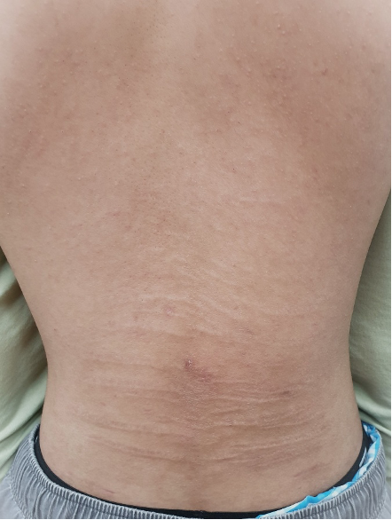
In 90% of cases (72 out of 80), the lesions were confined to the lower back, occasionally spreading to the mid-back or flanks. A few reports described atypical locations, including the face, thighs, knees, legs and upper limbs.
The lesions were usually yellowish or skin-coloured, though early ones sometimes appeared white, pink or red. They were palpable, slightly elevated and often detected incidentally during examination for unrelated complaints.
Histopathological findings consistently showed increased and thickened elastic fibres in the mid to deep dermis, arranged in parallel bundles or clumps, sometimes with irregular orientation or fragmentation, supporting a regenerative process rather than destruction of elastic tissue.
The etiopathogenesis of LFE remains uncertain, with none of the 37 articles providing a clear understanding. Most reports describe it as idiopathic, although two main theories have been proposed.

The first is the degenerative-regenerative hypothesis, which suggests that LFE results from abnormal elastic fibre repair following subclinical dermal damage or degeneration.
The second is the striae-distensae-related hypothesis, which considers LFE a regenerative counterpart or later stage of striae distensae, arising when overproduction of elastic fibres occurs after dermal stretching.
Evidence supporting this includes the coexistence of LFE and striae distensae in a small number of patients, and sequential evolution observed in a few others. Nevertheless, the two conditions remain clinically and histologically distinct. Striae distensae are typically atrophic, depressed and associated with conditions such as pregnancy, obesity or corticosteroid exposure, whereas LFE presents as palpable, thickened, elastotic bands without atrophy.
Approximately one quarter of documented adolescent cases were associated with recent growth spurts, and a small proportion were linked to vigorous physical activity, including swimming, running and bodybuilding, suggesting that mechanical stretching or microtrauma may stimulate abnormal elastic fibre regeneration.
Familial clustering was documented in only three reports, insufficient to infer a genetic component, the researchers wrote, adding that no systemic associations or underlying connective tissue diseases were consistently identified.
Because LFE is asymptomatic and benign, patients may not seek medical attention, contributing to its apparent rarity and under-recognition. Furthermore, misdiagnosis as striae distensae has been noted in several reports due to overlapping morphology, they said.
“However, LFE should be distinguishable from SD based on the latter’s clinical characteristics – morphology, typical sites of involvement [e.g., abdomen, gluteal region and proximal extremities], and associated risk factors [e.g., obesity, pregnancy or steroid use], as well as histological findings – elastolysis in the acute stage, with later stages exhibiting epidermal atrophy and densely packed, thin, horizontally arranged collagen bundles within the dermis.”
Therapeutic experience with LFE is limited and largely disappointing. Interventions such as topical and intralesional corticosteroids, topical retinoids, centella asiatica extract, and platelet-rich plasma have been tried in isolated cases and small series, with little or no clinical improvement.
The condition typically persists without progression or complications, and no malignant transformation has ever been reported.
The main clinical relevance of recognising LFE lies in differentiating it from other linear dermatoses rather than in initiating active treatment.
Dr Aung tells DR that he has only seen two cases of LFE in his career so far, but it could easily be mistaken for other conditions. Given there is no treatment identified to date, and it is unlikely the condition will spontaneously resolve, reassurance is the best advice.
He says the demographic trends derived from available data revealing that LFE affects males disproportionately and displays a bimodal age distribution, peaking in puberty and late adulthood was very interesting and definitely linked it with growth spurts.
There is a need for more research to understand the condition more, although so far there are no signs that it is a red flag for other diseases, says Dr Aung.
“As far as we know this condition is benign and not associated with any other underlying or serious conditions at all,” he says.
The researchers noted the limitations of the study, primarily the fact that the global literature comprises only 80 cases reported over three decades, nearly all from single or dual case reports or small institutional series.
No population-based or multicentre studies have yet been conducted, and the reliance on English-language publications introduced potential bias.
Data completeness also varied, with inconsistent reporting of age, sex, lesion duration and histopathological details.
However, taken together, the accumulated evidence positions LFE as an under-recognised, benign dermal condition that is most frequently observed in males, typically during adolescence or in later adulthood, they said.
The absence of effective treatment underscores the importance of accurate diagnosis and patient reassurance rather than therapeutic intervention.
For now, dermatologists are advised to keep LFE in mind when encountering horizontal linear lesions on the back, particularly in adolescent males or older adults.
The condition’s benign nature means that recognition, rather than treatment or even lengthy investigation and tests, remains the key to effective management.
